Radiology and imaging investigations
The importance of radiology and imaging investigations
The nowadays dentistry imaging investigations are indispensable in order to establish a correct diagnosis and developing a treatment plan. The type of investigation varies depending on the clinical situation and treatment to be performed. Imaging investigations are made both in the first visit, and later, during treatment period (intraoperative, postoperative, etc.), its completion and within the regular checks.
Types of imaging investigation
Our clinic is equipped with the high tech equipment with low levels of radiation, which uses the ALARA principle (As Low As Reasonably Achievable) according to which prevails obtaining high quality images with an as small as possible radiation dose, that can provide almost any type of imaging investigation required in the actual dental treatment.
- Intraoral digital X-rays (retroalveolar, bite-wing, complete dental status)
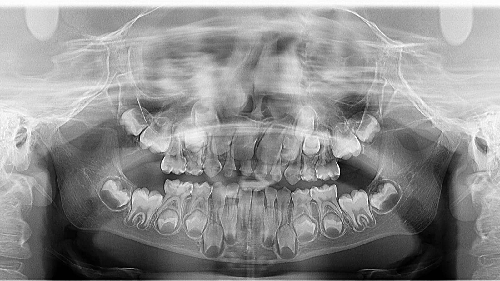
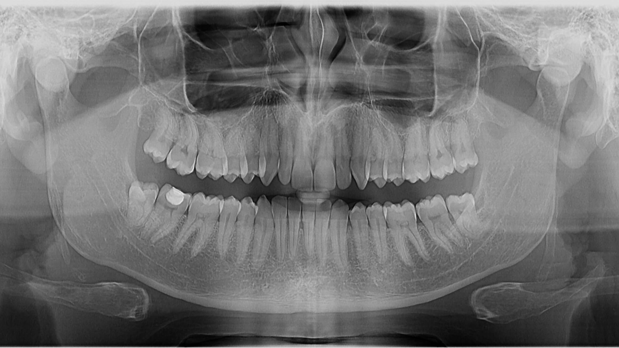
- Panoramic X-rays: 2D digital panoramic X-rays for children and adults;
- Panoramic X-rays for orthodontics;
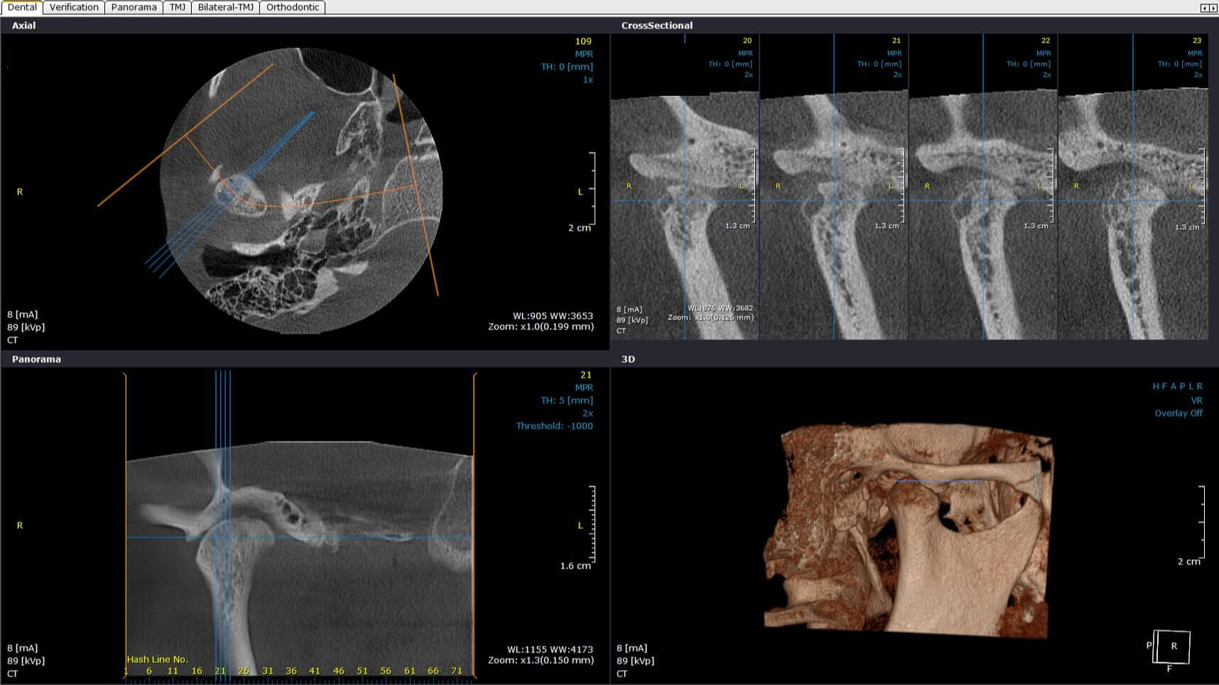
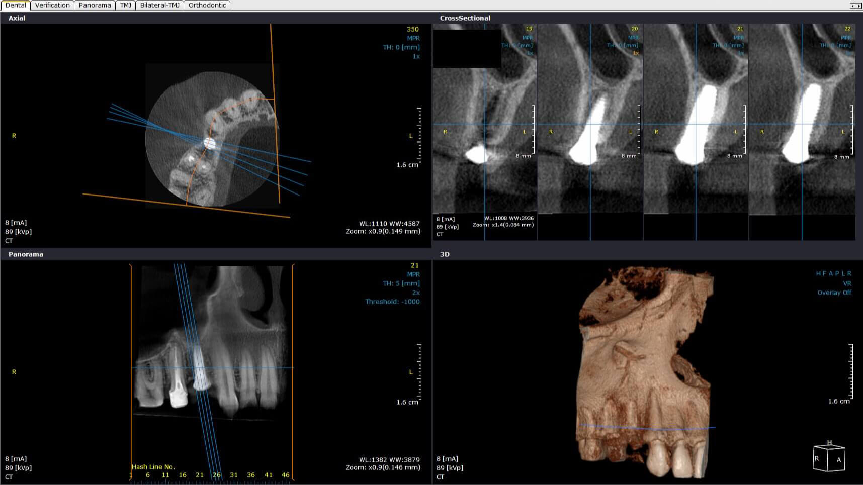
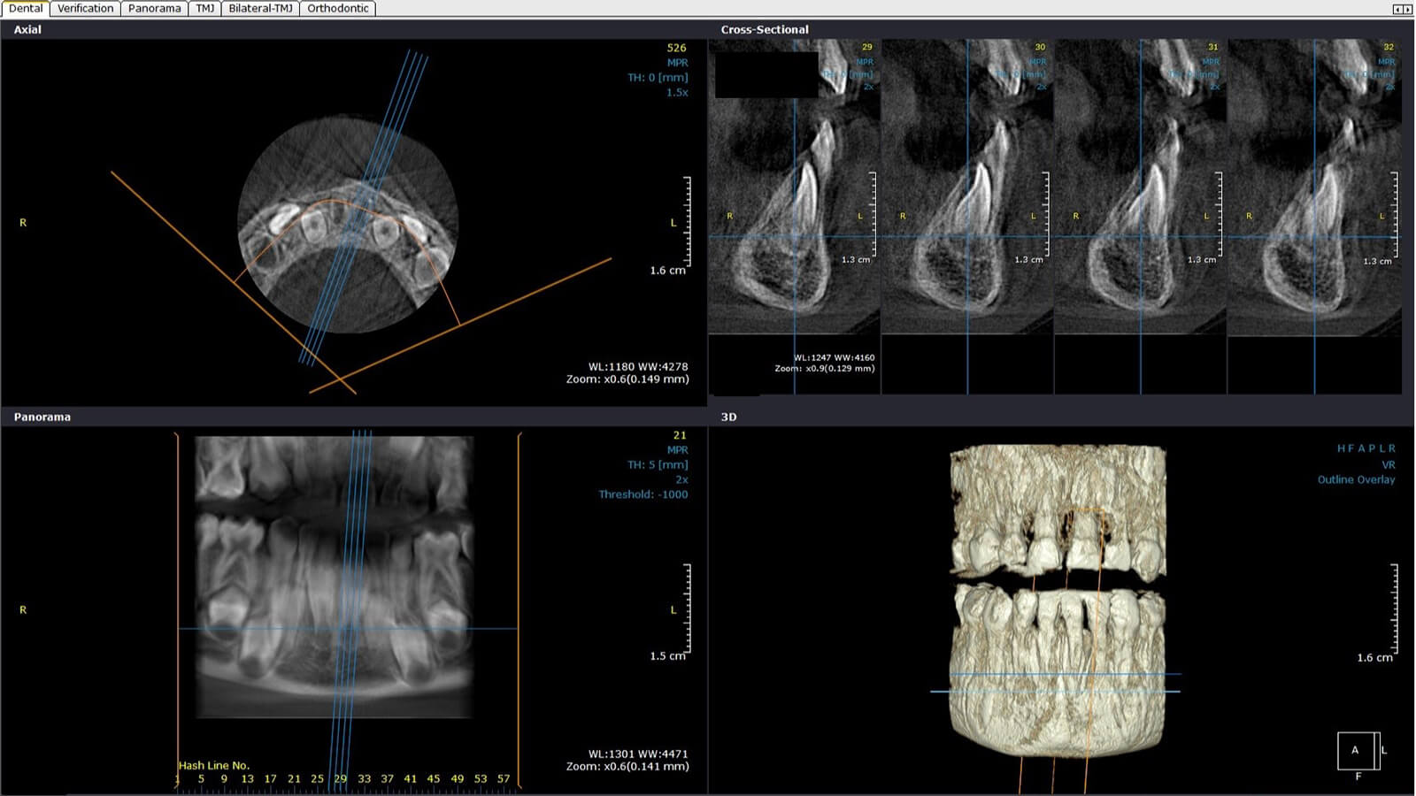
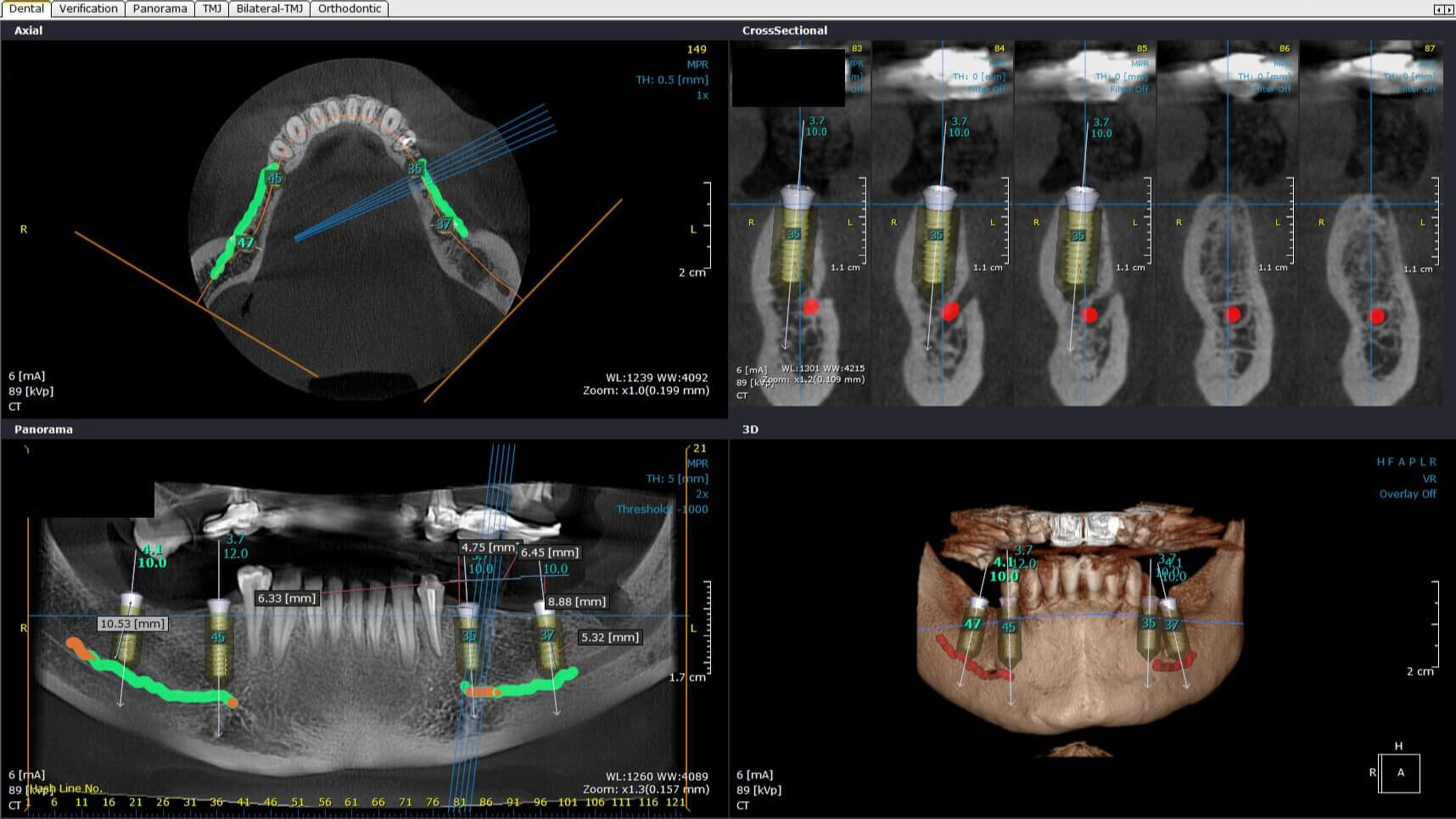
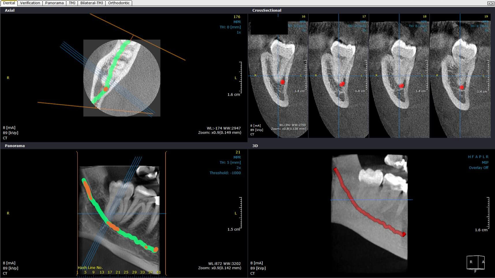
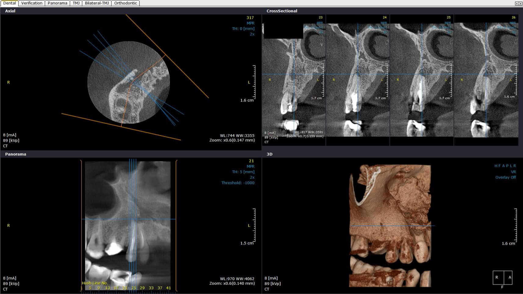
- 3D imaging tests: CBCT – Cone Beam Computed Tomography;
- Computed tomography of the temporomandibular joint (TMJ);
- 3D reconstruction and complex simulations of surgical and implant treatments.
What you should know about CBCT?
CBCT uses one small ray beam, it has a conical shape and specifically interest chosen by operator (scanning a group of teeth, an arcade, the whole tooth). The scanner needs only a few seconds and a single rotation around the patient’s head to get a good 3D image, in terms of quality. Using cone beam, the delimitation of interest area and short time of effective irradiation make the radiation dose in case of CBCT technology to be 10 to 20 times smaller than that used in the case of volumetric tomography (classic).
Photo gallery